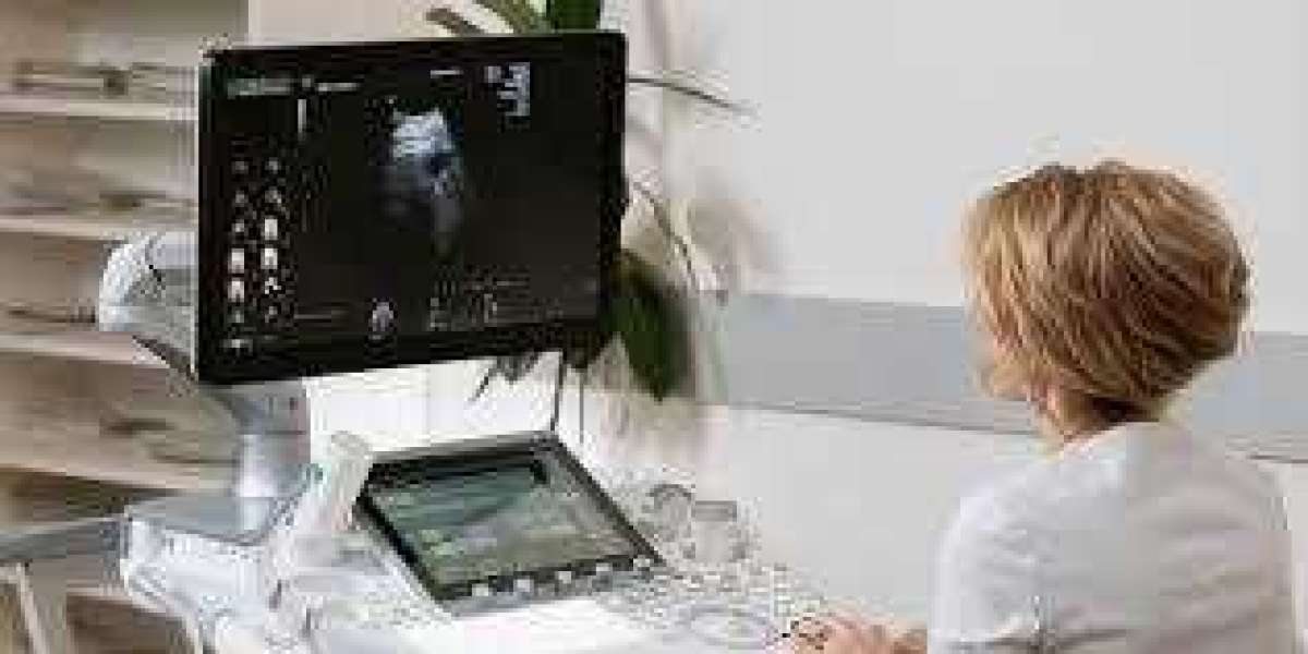
Ultrasound, also known as sonography, has become an essential component of modern medical diagnostics. Utilizing high-frequency sound waves to create images of internal structures, this technology offers a non-invasive, cost-effective, and widely available method for assessing various medical conditions. From prenatal care to the evaluation of organ function, ultrasound is transforming how healthcare professionals diagnose and treat patients.
The Science Behind Ultrasound
Ultrasound technology operates on the principle of sound wave propagation. When the transducer, a handheld device, is placed on the skin, it emits sound waves that penetrate the body. As these waves encounter different tissues, organs, and fluids, they bounce back to the transducer at varying speeds. A computer then interprets these returning sound waves, creating real-time images that can be visualized on a monitor.
The safety of ultrasound is rooted in its non-ionizing nature. Unlike X-rays or CT scans, which use potentially harmful ionizing radiation, ultrasound does not expose patients to radiation. This makes it a preferred choice for diagnostic imaging, particularly in sensitive populations such as pregnant women and infants.
Applications in Obstetrics and Gynecology
One of the most well-known applications of ultrasound is in obstetrics and gynecology. Ultrasound is widely used to monitor the health and development of a fetus during pregnancy. It allows healthcare providers to assess fetal growth, check for congenital abnormalities, and evaluate the position of the placenta. Doppler ultrasound, a specialized form of the technology, is used to measure blood flow in the umbilical cord, placenta, and fetal heart.
In addition to prenatal care, ultrasound is also invaluable in diagnosing gynecological conditions. It can help detect ovarian cysts, fibroids, and other abnormalities in the female reproductive system. As a non-invasive tool, ultrasound has largely replaced more invasive diagnostic methods such as exploratory surgery, reducing the risks associated with such procedures.
Cardiac Ultrasound: Echocardiography
Echocardiography, or cardiac ultrasound, is another critical use of this technology. It is employed to assess the structure and function of the heart. By capturing images of the heart's chambers, valves, and blood flow, echocardiography helps physicians diagnose and manage conditions such as heart failure, congenital heart defects, and valve disorders.
One of the major advantages of echocardiography is its real-time imaging capability, allowing doctors to monitor the heart as it beats. This makes it particularly useful in stress tests, where the heart's function can be evaluated under physical exertion. Furthermore, advances in 3D echocardiography provide detailed images, allowing for better surgical planning and more accurate diagnoses.
Abdominal Ultrasound: Assessing Organ Health
Abdominal ultrasound is widely used to evaluate the health of various internal organs, including the liver, kidneys, gallbladder, pancreas, and spleen. This type of ultrasound is instrumental in diagnosing conditions such as gallstones, liver disease, and kidney stones. Additionally, it is used to monitor the progression of chronic conditions like fatty liver disease or cirrhosis.
In emergency settings, abdominal ultrasound plays a crucial role in trauma care. Known as the FAST (Focused Assessment with Sonography for Trauma) exam, this quick, non-invasive procedure helps doctors detect internal bleeding or fluid accumulation in the abdominal cavity. The speed and accuracy of abdominal ultrasound make it a vital tool in life-threatening situations where rapid diagnosis is necessary.
Musculoskeletal Ultrasound: Visualizing Soft Tissues
Musculoskeletal ultrasound has gained prominence in diagnosing and managing injuries and conditions affecting the muscles, tendons, ligaments, and joints. It is often used to evaluate conditions such as tendonitis, muscle tears, and joint inflammation. Unlike MRI or CT scans, musculoskeletal ultrasound provides real-time imaging, allowing doctors to assess the movement of soft tissues as a patient moves.
This type of ultrasound is also used in guiding minimally invasive procedures such as joint injections or aspirations. By visualizing the needle's path in real time, physicians can accurately deliver treatment to the affected area, improving the safety and efficacy of the procedure.
Vascular Ultrasound: Monitoring Blood Flow
Vascular ultrasound is another critical application, used to assess the blood flow in arteries and veins. This form of ultrasound can detect blockages, blood clots, and aneurysms. It is particularly valuable in diagnosing deep vein thrombosis (DVT), a potentially life-threatening condition where blood clots form in the deep veins of the legs.
Doppler ultrasound, which measures the speed and direction of blood flow, is frequently used in vascular assessments. By evaluating how blood flows through vessels, healthcare providers can detect abnormal flow patterns, which may indicate blockages or narrowing of the arteries. This is especially important in diagnosing and managing peripheral artery disease (PAD), a condition that can lead to limb amputation if left untreated.
Advances in Ultrasound Technology
Recent advancements in ultrasound technology have expanded its diagnostic capabilities and improved image quality. Portable ultrasound devices, for example, are now available, allowing for point-of-care imaging in emergency rooms, operating theaters, and even remote locations. These devices offer the same diagnostic power as traditional ultrasound machines but in a compact, mobile form.
3D and 4D ultrasound are two innovations that provide enhanced imaging. While 3D ultrasound creates three-dimensional images of internal structures, 4D ultrasound adds the element of time, allowing for real-time visualization of movement. These advancements have proven particularly useful in obstetrics, where parents can see detailed, moving images of their developing baby.
Elastography is another emerging ultrasound technique that measures tissue stiffness. This can be used to assess the liver for fibrosis or tumors, providing a non-invasive alternative to biopsy. By quantifying tissue elasticity, elastography allows doctors to distinguish between benign and malignant lesions, potentially reducing the need for invasive diagnostic procedures.
The Role of Ultrasound in Guided Procedures
Ultrasound’s ability to provide real-time imaging has made it invaluable in guiding various medical procedures. In interventional radiology, ultrasound is often used to guide needle biopsies, catheter placements, and drainage procedures. By visualizing the internal structures, doctors can accurately place needles or instruments, minimizing the risk of complications.
In pain management, ultrasound-guided injections have become a common practice. By visualizing the target area, such as a joint or nerve, physicians can deliver medication precisely where it is needed, enhancing the effectiveness of the treatment.
Conclusion
Ultrasound has firmly established itself as a cornerstone of modern medical diagnostics. Its non-invasive nature, coupled with its ability to provide real-time, high-quality images, has made it a preferred tool across various medical fields. From obstetrics and cardiology to musculoskeletal and vascular assessments, ultrasound offers a versatile and safe method for diagnosing a wide range of conditions.
As technology continues to evolve, the applications of ultrasound are expected to expand further. With innovations such as portable devices, elastography, and 4D imaging, ultrasound will likely remain at the forefront of diagnostic medicine, offering increasingly precise and accessible healthcare solutions.


